(Horizontal Visible Iris Diameter)
SpecialEyes’ empirical fitting method using the SpecialEyes Arc Length Calculator takes into account corneal diameter, also known as horizontal visible iris diameter (HVID). If your patient appears to have a HVID that is larger or smaller than average (outside 11.6mm – 12.0mm), we advise measuring the patient’s HVID and incorporating this value into the initial design, as this will increase the probability that your first lens fitting will be successful.
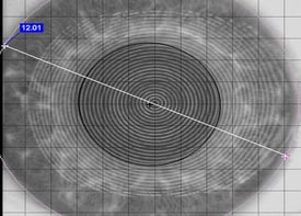
Technique 1: Corneal Topographer
Use the biometric ruler within the corneal topography software program to measure your patient’s horizontal visible iris diameter (HVID). It may be necessary to measure at a 45 degree oblique angle as the topographer may cut off part of the cornea in the horizontal plane. For accuracy and consistency, always measure from the white part of one side of the eye directly across to the white part of the other side of the eye. Reference the example image below; the corneal diameter or visible iris diameter measures 12.01mm.
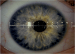
Technique 2: Slit Lamp & Reticle
Remove an eye piece from the slit lamp and insert the reticle. Use the slit lamp and integrated reticle to obtain a direct horizontal measurement of the patient’s visible iris diameter. Reference the example image below; the corneal diameter or horizontal visible iris diameter measures 11.8mm.
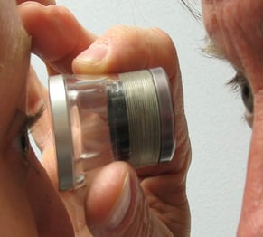
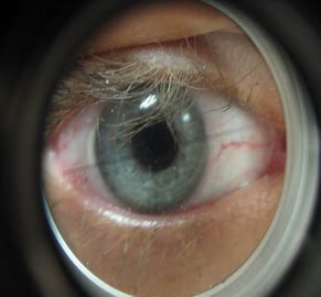
Technique 3: Loupe, 7X, Peak Scope
This device is commonly used for measuring the diameter of a GP contact lens, but can be an easy and efficient method to measure the horizontal visible iris diameter (HVID). Looking through the loop get close to the patient’s cornea; almost uncomfortably close similar to direct ophthalmoscopy. Use the measurement ruler built within the loop to measure the patient’s HVID as accurately as possible. Reference the example images below.